

심장이 갑자기 두근거리고, 소화가 안 되고, 밤에는 잠이 오지 않습니다. 병원을 몇 군데나 돌며 검사를 받았지만 결과는 늘 "정상", 돌아오는 말은 "신경성"이었습니다. 이렇게 여러 곳이 동시에 아픈데 원인이 보이지 않을 때 살펴봐야 할 것이 자율신경실조증입니다.
자율신경실조증 한눈에 보기
- 심장·호흡·소화·체온·수면을 자동으로 조절하는 자율신경의 균형이 깨져, 검사로는 이상이 없는데 여러 증상이 한꺼번에 나타나는 상태입니다.
- 일반인 10명 중 1명꼴로 이런 원인 모를 신체 증상을 겪고, 대개 여러 증상이 겹쳐 나타납니다.
- 오래 이어진 스트레스와 불안, 수면 부족, 감염 후 회복 지연이 대표적인 원인이며, 심박변이도 같은 검사로 자율신경 상태를 확인할 수 있습니다.
- 치료후기 71건
- 의료진 칼럼 28편
- 건강정보 17편
- 온라인 상담 9건
자율신경실조증이란?
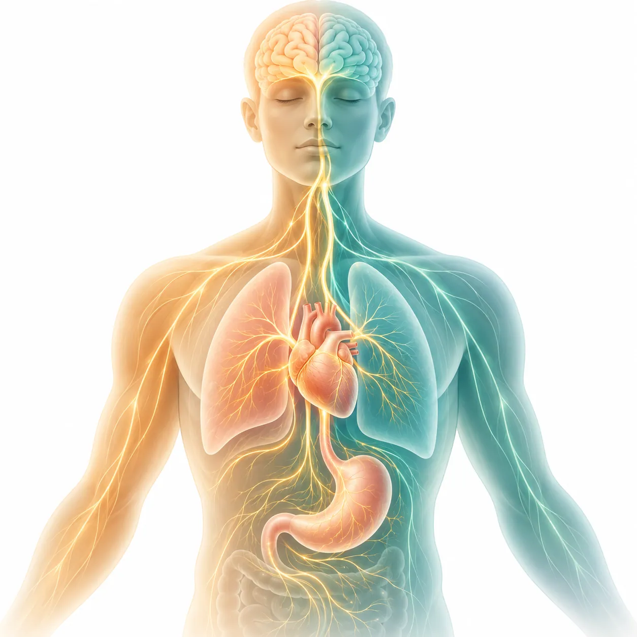
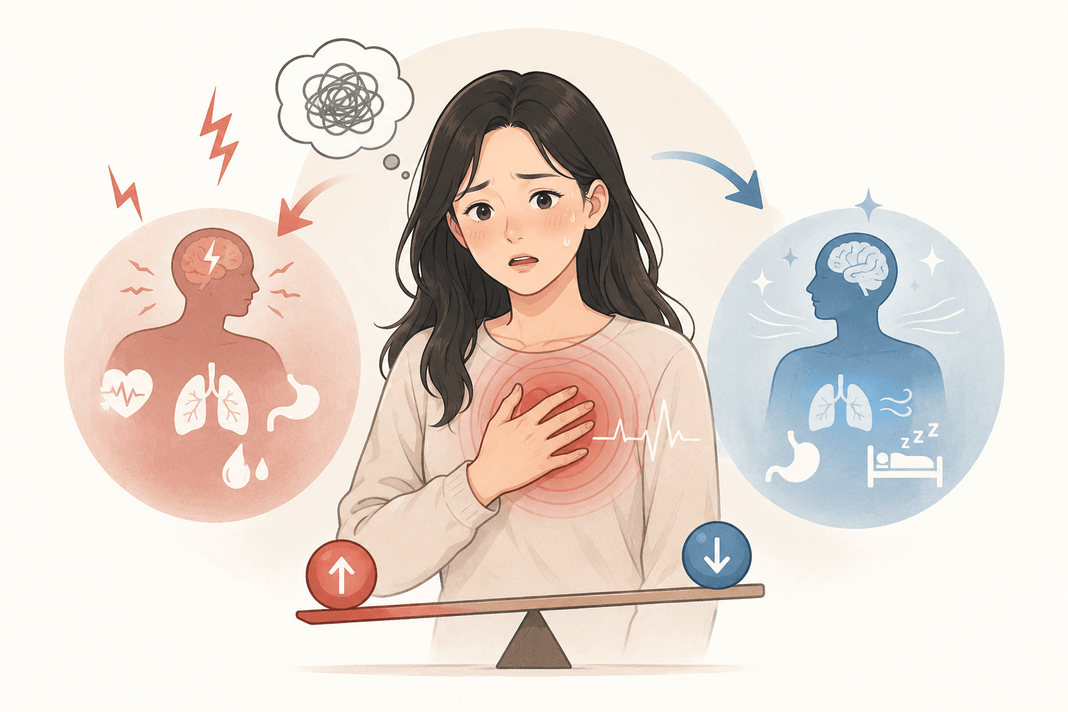
자율신경은 우리가 의식하지 않아도 심장 박동, 호흡, 소화, 체온, 면역을 조절하는 신경입니다. 몸을 긴장시키는 교감신경과 쉬게 하는 부교감신경이 시소처럼 균형을 맞추는데, 부교감신경의 중심인 미주신경은 기분과 면역, 소화와 심박을 함께 조절하며 장과 뇌를 잇는 통로 역할도 합니다[1]. 이 균형이 오랫동안 한쪽으로 기울어 제자리를 찾지 못하는 상태를 흔히 자율신경실조증이라고 부릅니다.
자율신경실조증은 한 가지 병명이라기보다 "검사로는 원인이 드러나지 않는 여러 신체 증상"을 묶어 부르는 말에 가깝습니다. 덴마크 일반인 조사에서는 이런 기능성 신체 증상군이 10명 중 1명꼴(9.3~10.7%)로 나타났고, 과민성대장·만성피로·만성통증이 서로 겹쳐 한 가지만 있는 경우는 드물었습니다[2]. 유럽 연구진은 이런 상태를 "몸만의 병도, 마음만의 병도 아닌" 뇌와 몸의 상호작용 문제로 보자고 제안합니다[3].
이런 증상이 있나요?
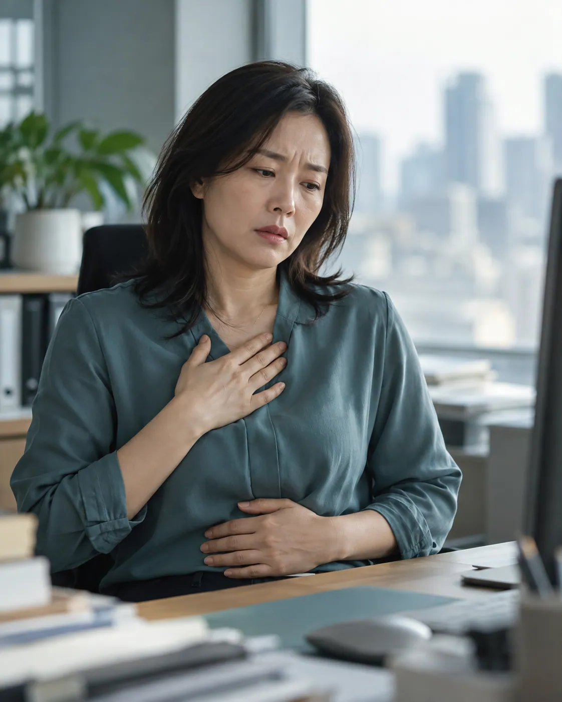
- 가만히 있어도 심장이 두근거리거나 철렁 내려앉는다
- 가슴이 답답하고 숨이 끝까지 쉬어지지 않는다
- 조금만 먹어도 더부룩하고, 긴장하면 배가 아프거나 설사를 한다
- 손발이 차고, 긴장하면 손이나 겨드랑이에 땀이 많이 난다
- 머리가 무겁고 어지러우며, 뒷목이 자주 뻣뻣하다
- 피곤한데 잠이 오지 않거나, 자도 개운하지 않다
- 병원 검사를 여러 번 받았지만 "이상 없음"이라는 말만 들었다
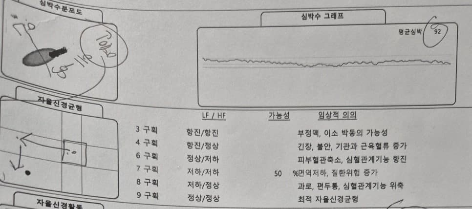
서로 다른 부위의 증상이 두세 가지 이상 함께 있고 검사에서는 원인이 나오지 않았다면 자율신경의 균형을 확인해 볼 필요가 있습니다. 심박변이도(HRV)는 심장 박동 간격의 미세한 변화를 분석해 교감·부교감신경의 균형을 보는 검사로, 스트레스를 받으면 부교감신경 활동이 떨어지는 쪽으로 변합니다[4].
자율신경실조증의 표준 치료
자율신경실조증으로 병원을 찾으면 보통 두근거림에는 심장약, 소화불량에는 위장약, 불면에는 수면제처럼 증상마다 약을 받게 됩니다. 증상은 잠시 가라앉아도 균형이 깨진 바탕은 그대로라 약을 끊으면 다시 시작되기 쉽습니다. 그래서 증상을 하나씩 누르기보다 자율신경의 균형 자체를 되찾는 방향으로 접근해야 합니다. 이를 위한 전문적인 자율신경치료가 따로 있고, 꾸준한 운동도 도움이 됩니다. 실제로 태극권·요가 같은 심신 운동을 꾸준히 한 사람은 심박변이도가 개선되고 스트레스 수준이 뚜렷하게 낮아졌습니다[7].
해움한의원은 이렇게 치료합니다

해움한의원은 검사로 자율신경 상태를 확인하고, 한약·약침·상담을 아우르는 자율신경치료로 균형을 되찾도록 돕습니다.
1단계. 자율신경 상태를 검사로 확인합니다
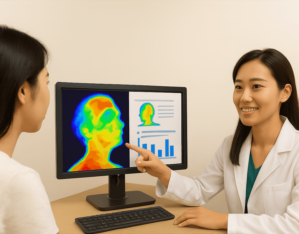
심박변이도(HRV)로 교감·부교감신경의 균형을, 뇌파로 긴장과 각성 상태를, 적외선 체열검사(DITI)로 몸의 순환과 열 분포를 확인합니다. 검사 결과와 함께 증상이 시작된 시기, 스트레스와 수면, 생활 습관을 자세히 살펴보고 원인을 정리합니다.
2단계. 긴장을 풀고 회복력을 되살립니다
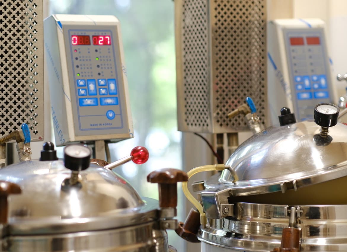
체질과 상태에 맞춰 한약을 짓습니다. 약침·도침·추나로 목·등·가슴의 긴장을 풀어 자율신경이 지나는 길을 편하게 합니다.
3단계. 마음과 생활까지 함께 다룹니다
심리상담사 1급 자격을 갖춘 원장이 스트레스의 원인과 생각 습관을 함께 살핍니다. 호흡법, 수면 습관, 몸을 움직이는 방법처럼 일상에서 부교감신경을 깨우는 방법을 안내해, 치료가 끝난 뒤에도 균형을 지킬 수 있게 돕습니다.
4만 건의 처방 사례가 말해주는, 한방 맞춤 치료의 힘


자율신경실조증 치료후기 총 71건
환자분들이 남긴 치료후기입니다. 본문은 로그인 후 보실 수 있습니다.

스트레스로 인한 자율신경 문제가 불러온 화병, 불면증
2024. 06. 28.
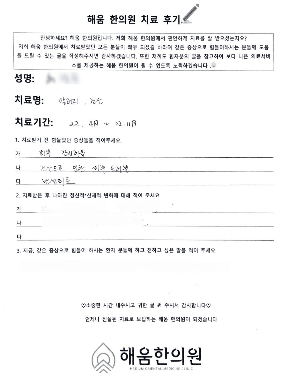
건선치료제로 효과 없는 피부건선, 한약 6개월 치료 사례.. 자율신경이 면역과 건선 혈액순환에 미치는 영향
2023. 01. 17.
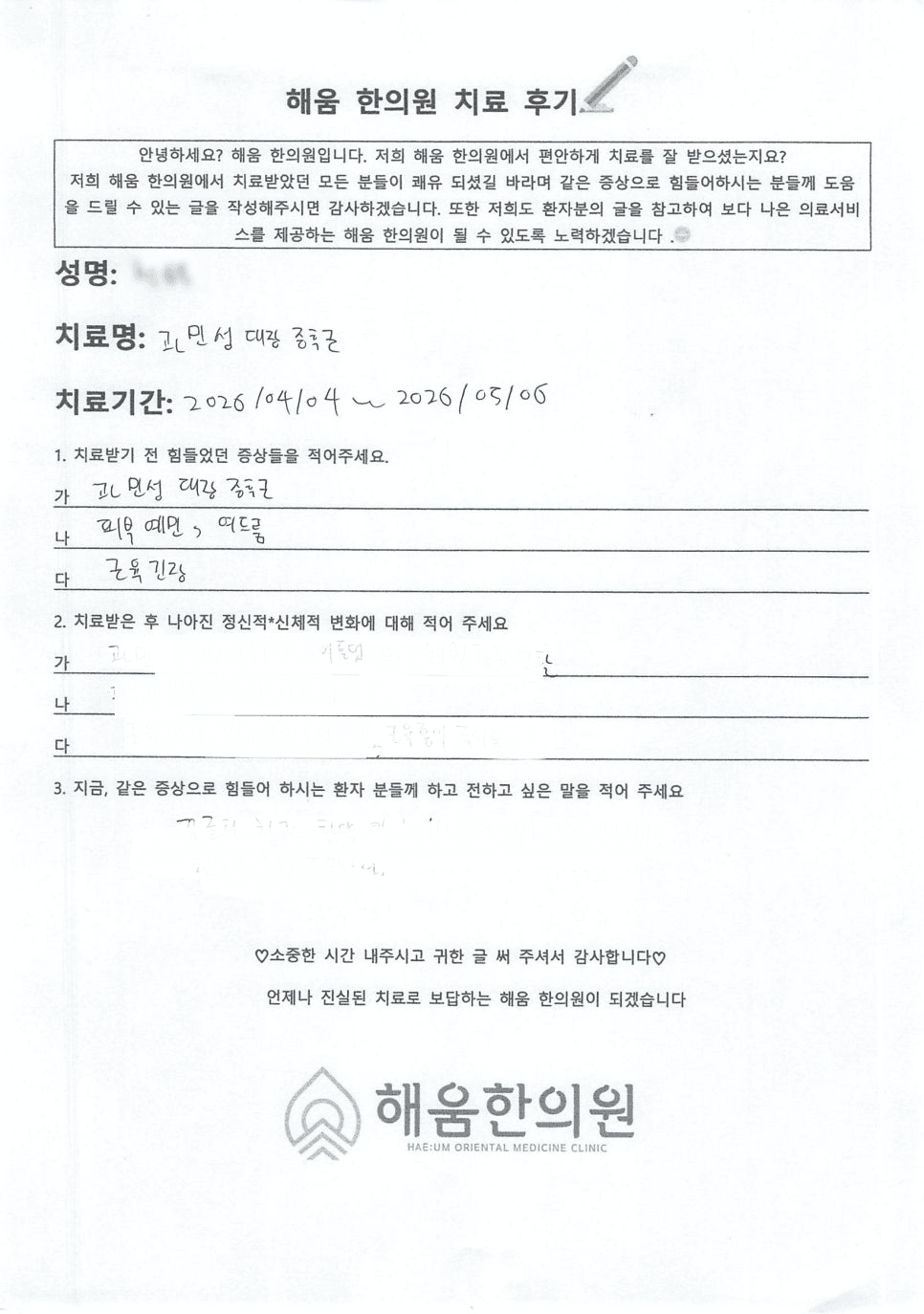 관련 후기
입대 전 갑자기 발생한 과민성대장증후군, 한의원에서 치료가 될까요?
2026. 06. 01.
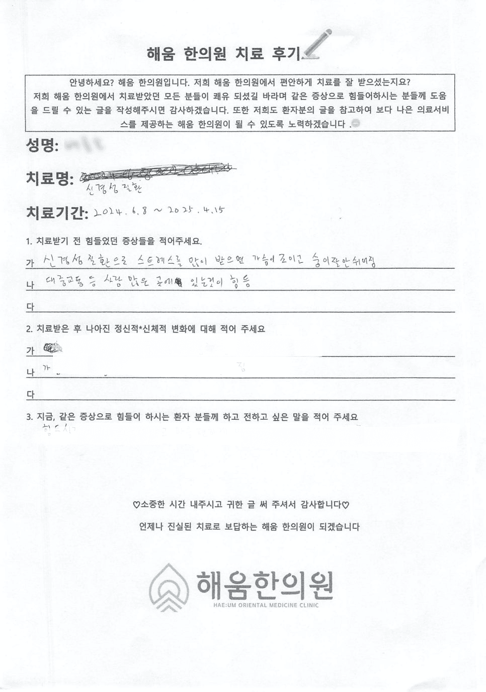 관련 후기
갑자기 화장실을 못참겠다면? 과민성대장증후군가스잦은설사 한약으로 치료가 될까요? 과민성대장증후군병원
2026. 05. 27.
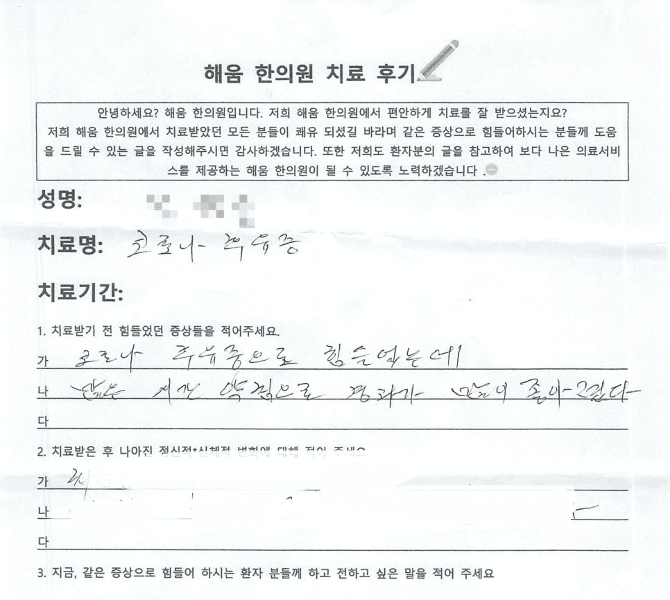 관련 후기
코로나 백신 맞고 기억이 안날 때는? 머리가 멍해요. 코로나 브레인포그 후유증 브레인포그 극복 혈관성 치매 한의원 후기
2026. 05. 04.
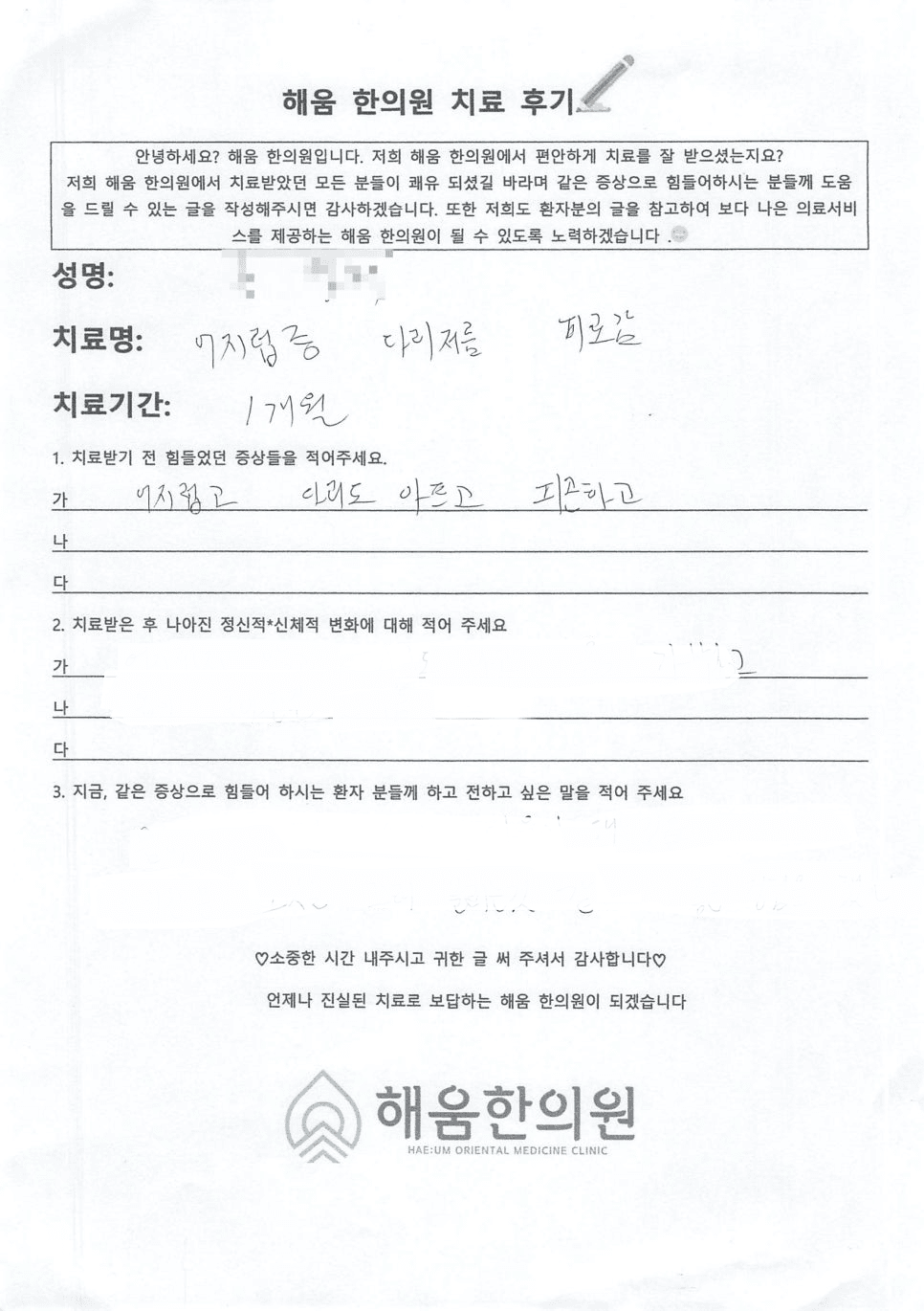 관련 후기
갑자기어지럽고식은땀이 흐른다구요?
2026. 04. 25.
다른 증상으로 내원하신 분들의 후기 중 ‘자율신경실조증’이 증상으로 함께 언급된 기록
- 치아가 욱씬거렸던 이유는 뭐였을까요?? 3차신경통치료 알아보기2026. 04. 16.
- 재수생에게 갑자기 찾아온 스트레스심장두근거림과 급체이유는 뭘까요?2026. 04. 14.
- 두통 속울렁거림 식은땀, 단순 소화불량이 아니라고요?2026. 04. 09.
- 공황장애 유전되나요? 공황장애2026. 02. 12.
- 목칼칼기침 독감후잔기침,숨참 으로 찾아오신 환자분2026. 02. 02.
- 명치답답 소화불량, 담적 증상 같은데 원인이 없대요. 정신과 문제인 걸까요?2026. 01. 31.
- 갱년기인 줄 알았는데 화병이었습니다 발후끈거림 홧병2026. 01. 30.
- 건선치료제로 효과 없는 피부건선, 한약 6개월2026. 01. 28.
- 시험만 보려고 하면 배가 아프고 설사하고 심장이 두근거려요2026. 01. 19.
- 심장이 철렁하고 가슴이 아파요..심장뻐근함도 한약으로 치료가 되나요?2026. 01. 12.
나머지 55건 더 보기접기
- 검사는 이상 없는데 복통이 심해요..제가 예민한 걸까요?2026. 01. 09.
- 직장 스트레스와 과로로 악화된 편두통·삼차신경통 환자2026. 01. 06.
- 소화불량두통, 체할까 봐 항상 조심스러웠던 마음이 이제는 편해졌어요2026. 01. 05.
- 극심한 생리통, 알고보니 자궁물혹이래요.. 아랫배 빵빵 생리통 심해짐, 한약으로 생리통 없어짐 가능할까요?2025. 12. 27.
- 검사에선 이상 없다는데, 명치가 왜 계속 아플까요? 소화불량 명치통증 내시경 원인 치료2025. 12. 16.
- 이건 붓기일까요 살일까요? 얼굴살 팔뚝살 빼기 상체 비만 다이어트2025. 07. 21.
- 명치끝 통증, 알고보니 자궁내막암 후유증이었다고?2025. 07. 08.
- 기침 콧물 멈추는법? 목동 한의원에서 알려주는 코비염 해결법2025. 02. 15.
- 갑상샘항진증 아급성갑상선염 치료 환자, 피로감 소화불량 고지혈증 완화 한약 2달간2025. 01. 25.
- 목넘김이물감 매핵기증상 호흡곤란2024. 12. 28.
- 공황장애 가슴통증 가슴답답 두근거림, 목디스크, 어깨결림 증상, 불면증 1개월2024. 12. 13.
- 코로나 백신 후유증으로 인한 공황장애 발작2024. 06. 28.
- 6개월간 생리를 안해서 내원하신 생리불순2024. 06. 08.
- 이유모를 공황장애 한약으로 치료한 후기2024. 06. 07.
- 치료 안 돼서 고생하던 과민대장증후군2024. 06. 06.
- 불면증두통, 호흡곤란, 다이어트와 함께2024. 05. 24.
- 심한 변비 배아픔에 직빵, 소화불량 불안 불면 3개월2024. 05. 02.
- PTSD 외상후스트레스장애 증상으로 찾아온 초등학생, 한약 치료로 두근거림,호흡곤란,불안장애,소화불량이 좋아졌어요2024. 05. 01.
- 코로나로 악화된 이명 코로나후유증 한약 6개월2024. 04. 21.
- 안하던 생리를 3일만에 시작했어요! 생리통 생리불규칙 무월경 몸이 가뿐해졌다는 환자 사례2023. 12. 23.
- 아내와 사별후 찾아온 갑자기 가슴두근거림 손발저림 어지러움증2023. 12. 22.
- 생리전증후군인가요? 진통제를 하루 8알씩.. 심각한 생리통 편두통 변비 한약 먹고 좋아진 사례2023. 12. 13.
- 이노시톨부작용 여드름으로 고생하던 다낭성증후군 PCOS 환자, 한약 치료로 3개월만에 해결2023. 12. 12.
- 복통 설사가 너무 심해서 출근도 못하는 과민성대장 환자2023. 12. 10.
- 홧병으로 두근거리고 심장저림까지 있던 환자분 사례2023. 12. 09.
- 코로나로 악화된 공황장애 분열정동장애 한약 6개월2023. 11. 22.
- 스트레스인지 코로나후유증인지.. 왼쪽가슴통증손저림 만성피로 염증 한 방에!2023. 01. 18.
- 과민성대장증후군 불면증치료법 중에 한약이 최고?!2023. 01. 14.
- 풀어지는 변, 과민대장증후군 안녕~ 한의원에서 한약으로2023. 01. 11.
- 심장이 철렁, 심장두근거림이 너무 심하고 가슴통증까지 와요2023. 01. 10.
- 3개월간 고생한 옆구리통증 명치통증 호흡곤란 소화불량 1달만에2022. 12. 29.
- 스트레스성 공황장애 초기증상 한약 치료사례: 공황발작과 공황장애의 차이2022. 12. 27.
- 가슴두근거림,무기력,우울증,체력저하 한약 치료기2022. 06. 27.
- 소화불량으로 3개월동안 10kg 빠져서 고생하신 환자분 한약 치료기2022. 05. 12.
- 스트레스 받으면 화장실로 뛰쳐나가게 하는 복통설사2022. 05. 11.
- 토끼 같이 깜짝깜짝 잘 놀라는 친구의 불안장애 한약치료사2022. 04. 18.
- 스트레스성 폭식으로 급찐 살 9kg 감량!! 다이어트 성공 사례2022. 04. 04.
- 불면증 한약으로도 치료되나요? 말똥말똥 잠이 안 와서 다음날이 걱정되던 환자2022. 04. 02.
- 공황장애와 공황발작의 차이? 두근거림,이명,두통,불안감 한의원 1달2022. 03. 21.
- 알러지 체질 고칠 수 있나요? 한랭두드러기와 비염 한약 3개월2022. 03. 19.
- 만성피로,소화불량,불면.. 사는게 피곤하셨던 환자분 보약 먹고 기운 차리신 얘기2022. 03. 09.
- 잔병치레를 달고 살던 환자분, 한약으로 소화불량 위경련 예민함 치료를 통해 좋아진 사례2022. 03. 07.
- 선생님 점쟁이세요...? 숨쉴때 답답하고 가슴 통증에 두근거림까지 있던 환자2021. 12. 21.
- 공황장애도 전조증상이 있을때 예방해야해요 호흡곤란,식은땀,불안감 등 공황발작2021. 11. 24.
- 배가 계속 아프다고 하는 4살 아가, 내시경 관장 위장약 다 시도해도 소용없던 복통 다한증과 함께 좋아진 사례2021. 10. 29.
- 응급실 다녀온 후 생긴 식사불가,호흡곤란,불면 으로 직장을 그만둘뻔했던 분 완치 사례2021. 10. 29.
- 애기때부터 시작한 만성변비와 여드름 치료사례, 장건강이 피부건강에 얼마나 중요하냐면요2021. 10. 28.
- 야간근무로 인해 살찌고 감정기복이 너무 심했던 간호사 환자, 한 포 먹고 식욕이 뚝 떨어지고 마음이 편안해졌다고..2021. 10. 27.
- 10kg 감량! 스트레스로 인한 체중증가, 한약으로 화병과 비만 둘다 관리할 수 있답니다2021. 10. 15.
- 정신과약부작용으로 생긴 불안장애, 두근거림, 불면증 1달만에 거의 사라진2021. 10. 11.
- 숨이 쉬어지지 않는 가슴통증 한 번에 완쾌한 사례2021. 09. 08.
- 코로나후유증으로 조금만 움직여도 숨차고 기침이 심했던 사례2021. 08. 28.
- 내시경상 원인을 모르는 소화불량 약침 추나치료 3번만에 완쾌한 후기2021. 07. 16.
- 과민성대장증후군 시험볼때 쥐어짜는 복통으로 뛰쳐나가야했던 여학생 한약 치료후기2021. 07. 15.
- 코로나 백신 후유증으로 생긴 불면, 불안, 소화불량, 이명2021. 01. 14.
자율신경실조증, 누가 진료하나요?
담당 의료진
대표원장 박재은

“병이 아니라 그 뒤의 사람을 봅니다”
경희대학교 한의과대학에서 석사·박사 과정을 마쳤습니다. 심리상담사 1급 자격과 4만 건 이상의 한약 처방 경험을 바탕으로 진료합니다.
의료진 소개 더보기자율신경실조증, 자주 묻는 질문
자율신경실조증은 정식 병명인가요?
자율신경 상태를 검사로 확인할 수 있나요?
병원 약을 먹고 있는데 한방치료를 같이 받아도 되나요?
치료 기간은 얼마나 걸리나요?
생활에서 자율신경을 돌보는 방법이 있나요?
한의원 진료 기록이 남으면 불이익이 있나요?
해움한의원에서 자율신경실조증을 진료하나요?
자율신경실조증으로 내원하려면 어떻게 하나요?
자율신경실조증에 관해 알아두면 좋은 정보
17건자율신경실조증과 관련해 정리한 건강정보입니다.
배고플때손떨림 왜 생길까? 저혈당과 자율신경의 숨은 관계
배가 고픈데 왜 손까지 떨리는 걸까요?아침을 거른 날, 바쁜 업무 때문에 점심시간을 놓친 날, 혹은 다이어트 중 식사량을 줄인 날 갑자기 손이 떨리는 경험을 해보신 적이 있으신가요?처음에는 "배가 많이 고픈가 보다"

왼쪽 오른쪽가슴답답 원인, 심장일까? 자율신경일까?
“한쪽이던 답답함이 양쪽으로 번졌다면, 몸이 보내는 신호입니다”처음에는 가볍게 느껴집니다.왼쪽 가슴이 조금 답답한 느낌, 숨이 살짝 막히는 듯한 느낌.하지만 시간이 지나면서오른쪽까지 답답해지고,심지어는 가슴이 꽉 조

뒷목뻐근 두통 어지러움, 자율신경균형이 핵심입니다
“그냥 두통이겠지…” 하고 넘기고 계신가요?뒷목이 뻐근하면서 머리가 지끈거리고, 때로는 어지러움까지 동반되는 경험. 한 번쯤은 있으셨을 거예요.특히 스트레스를 받는 날이면목이 딱딱하게 굳고, 머리가 무겁고, 눈앞이

두근거림·호흡곤란 반복된다면, 공황장애 해결의 답은 자율신경입니다
“이제 밖에 나가는 것도 무서워요…”갑자기 숨이 가빠지고, 어지럽고, 심장이 미친 듯이 뛰는 느낌.이런 증상이 한 번이 아니라 반복되기 시작하면단순한 스트레스라고 넘기기 어려워집니다.특히∙ 사람이 많은 곳∙ 지하철,

갑자기속울렁 반복된다면? 담적병과 자율신경 문제를 살펴보세요
이유 없이 속이 울렁거릴 때, 몸은 이미 신호를 보내고 있습니다멀쩡하게 일상을 보내다가 갑자기 속이 울렁거리는 경험을 해보신 적 있으신가요?음식을 잘못 먹은 것도 아닌데 메스껍고, 속이 뒤집히는 느낌이 들며 심한 경

머리가아프고어지러워요, 두통과 어지럼증 반복된다면 자율신경을 확인하세요
진통제를 먹어도 낫지 않는 두통, 이유를 찾지 못해 답답하셨나요?머리가 무겁고 어지럽습니다.가만히 있어도 멍한 느낌이 들고, 심할 때는 속이 울렁거리거나 중심을 잡기 어려울 정도로 어지럽습니다.처음에는 피곤해서 그런
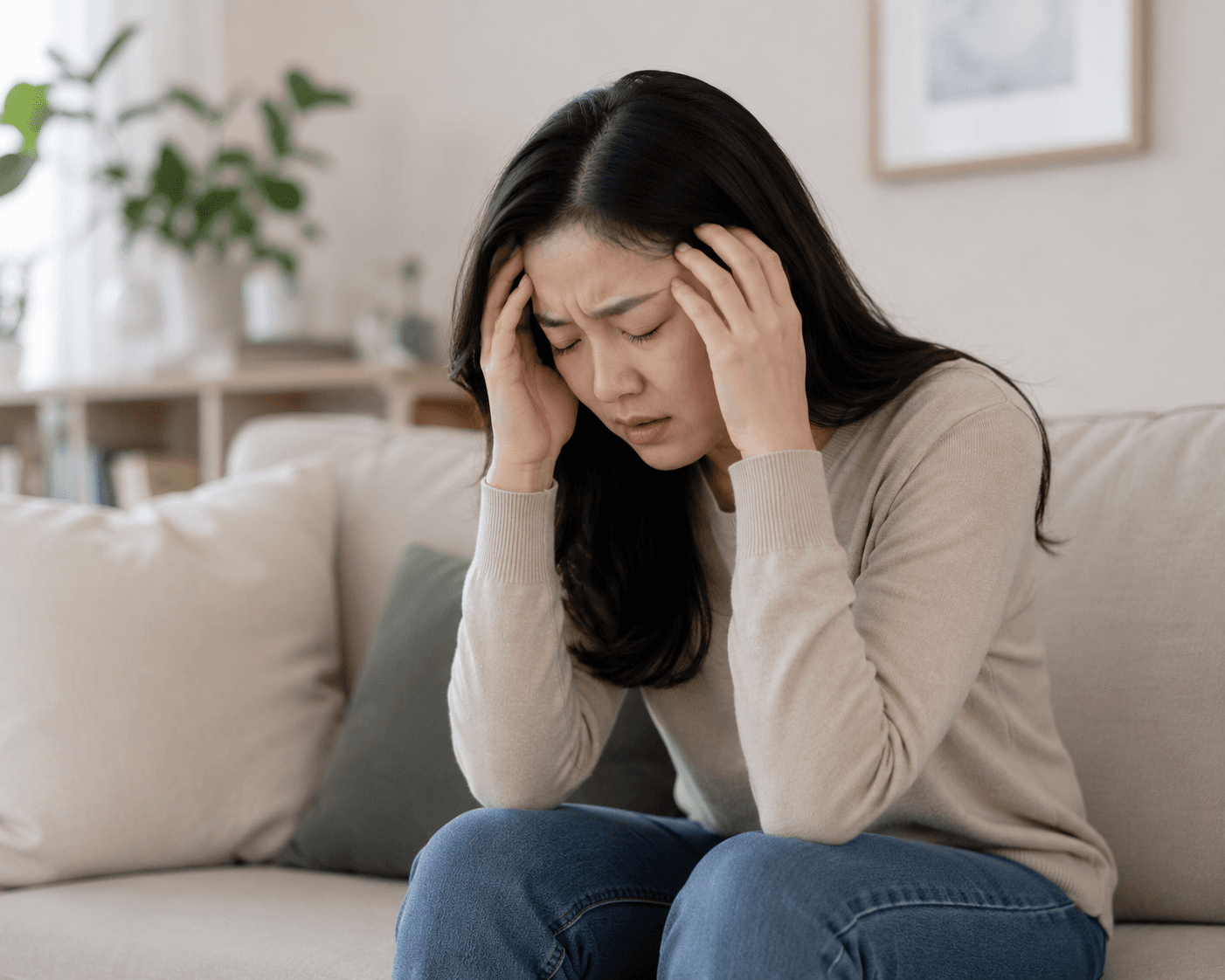
교감신경항진증 치료, 원인부터 바로잡아야 자율신경실조증에서 벗어날 수 있습니다
이유를 알 수 없는 가슴 두근거림, 혹시 교감신경이 과하게 흥분한 상태일까요?가슴이 갑자기 두근거리고 숨쉬기가 답답합니다.밤에는 쉽게 잠들지 못하고, 잠이 들어도 자주 깨서 피곤함이 계속됩니다.병원을 찾아 여러 검사
병원 검사에서는 정상인데 힘들다면? 자율신경실조증검사후기로 알아보는 원인
병원을 다녀도 이유를 모르겠다면최근 들어 이유를 알 수 없는 다양한 증상으로 의료기관을 찾는 분들이 크게 늘고 있습니다.머리가 아프고 어지럽습니다.가슴은 이유 없이 두근거립니다.식은땀이 흐르고 손발이 저립니다.입이
왼쪽가슴답답 반복된다면, 스트레스와 자율신경 불균형을 살펴보세요
심장은 괜찮다는데 왜 계속 답답할까요?왼쪽 가슴이 꽉 조이는 듯 답답합니다.숨을 크게 쉬어도 시원하지 않고, 밤에는 그 답답함 때문에 잠이 잘 오지 않기도 합니다.처음에는 심장 문제일까 걱정되어 심전도 검사나 심장

두근거림부터 소화불량까지... 모두 자율신경계이상질환 때문일 수 있습니다
검사에서는 이상이 없다는데, 몸은 계속 불편하신가요?머리는 무겁고, 심장은 이유 없이 두근거립니다.조금만 움직여도 어지럽고, 소화는 잘되지 않으며 잠도 깊게 들지 못합니다.병원을 찾아 심장검사와 혈액검사, 위내시경,

원인을 찾지 못한 여러 증상, 자율신경실조증치료법이 필요한 이유
이유 없이 아픈 곳이 늘어나는데 검사에서는 정상이라고 한다면머리도 아프고, 가슴도 답답하며 숨쉬기조차 불편합니다.소화도 잘되지 않고 어지러움까지 반복되는데 병원에서 여러 검사를 받아봐도 특별한 이상이 없다는 이야기를

홧병약 복용에도 낫지 않는다면 자율신경 균형을 확인해보세요
억눌러온 감정이 몸으로 나타나는 경우가 있습니다"가슴이 답답하고 한숨만 나와요.""화를 참다 보니 몸이 먼저 아파졌어요.""홧병약을 먹어도 크게 달라지는 게 없어요."이처럼 오랫동안 감정을 참고 살아온 분들 가운데

명치끝통증 원인, 위장 문제와 자율신경 균형을 함께 봐야 하는 이유
명치가 콕 아플 때, 그냥 체한 걸까요?명치 끝이 콕 찌르듯 아프거나,타는 듯 쓰리고 답답한 느낌이 들면대부분 먼저 “체했나?” 하고 생각합니다.그런데 명치 끝통증은단순 소화불량만으로 나타나는 증상이 아닐 수 있습니

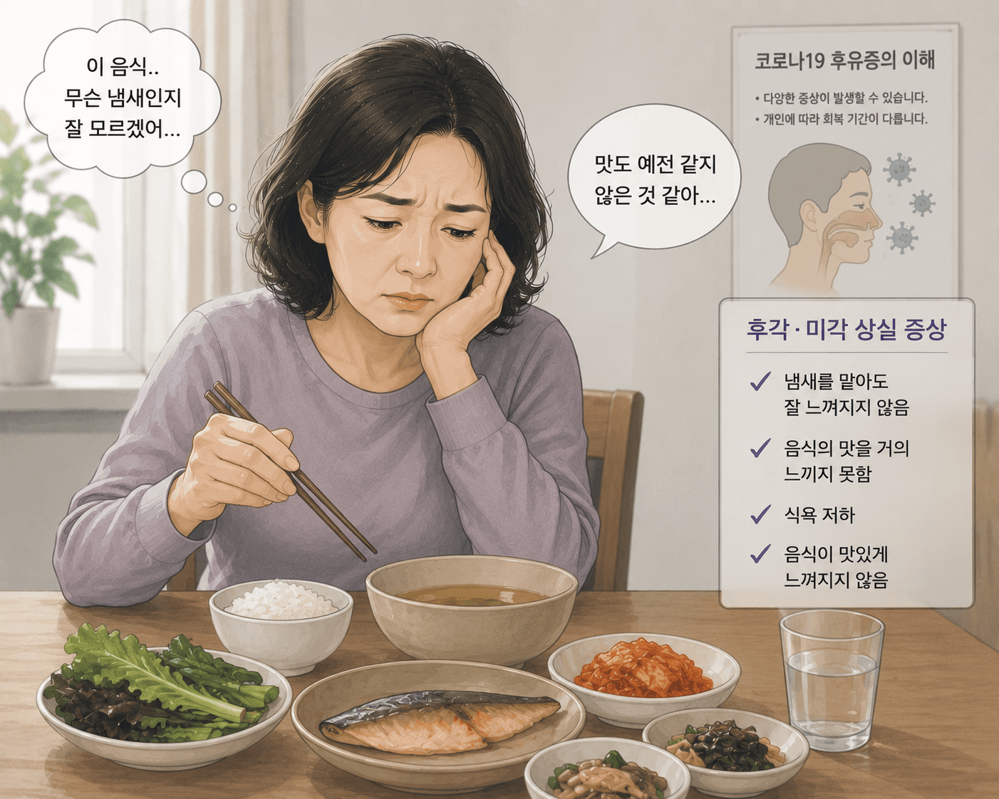
코로나후유증후각상실, 미각까지 사라졌다면 자율신경도 함께 확인해야 합니다
코로나는 끝났는데 냄새와 맛은 아직 돌아오지 않았나요?코로나19가 유행하던 시기보다 일상은 많이 회복되었습니다.하지만 완치 판정을 받은 뒤에도"커피 향이 전혀 안 느껴져요.""김치 맛도 맹물 같아요.""음식이 맛이
입안쓴맛과 건조함, 자율신경과 면역의 문제일 수 있어요
아무것도 먹지 않았는데 계속 쓴맛이 난다면아침에 일어났는데 입안이 텁텁하고 쓴맛이 올라올 때,혹은 하루 종일 아무것도 먹지 않았는데도 입에서 쓴맛이 맴도는 경험 있으신가요?“입안의 쓴맛은 단순한 입 문제라기보다 몸

조금만먹어도배부름, 소화 문제가 아니라 자율신경 문제일 수 있습니다
“분명 많이 먹지 않았는데, 왜 이렇게 답답할까요?”“밥을 몇 숟갈 안 먹었는데 벌써 배가 꽉 찬 느낌이에요.”“조금만 먹어도 더부룩하고, 소화가 안 되는 것 같아요.”혹시 이런 경험, 반복되고 있지 않으신가요?처음

반복되는위염, 자율신경이 무너지면 생기는 신호입니다
“괜찮아질 줄 알았는데, 왜 계속 반복될까요?”“속이 쓰리고, 밥을 먹으면 더부룩하고… 공복에는 또 아파요.”이런 증상 때문에 병원을 찾고 약을 먹어보셨나요?그런데도 이상하게 며칠 괜찮다가 다시 반복되는 경험, 해보

자율신경실조증, 질문 있어요
9건환자분들이 남긴 온라인 상담 질문과 의료진 답변입니다.
- 어지럼증이랑 심장 두근거림이 같이 나타나는데 자율신경 문제일까요ㅜ?2026. 10. 30.
- 자율신경실조증 치료를 여러 번 받은상태인데도 증상이 반복되는 중...2026. 10. 19.
- 자율신경계 질문있습니다2026. 05. 11.
- 자율신경계 검사를 받아야 할것같아요2026. 05. 06.
- 자율신경실조증검사2026. 04. 29.
- 자율신경실조증2026. 04. 09.
- 자율신경실조증 진단받고 약복용중인데 전혀 괜찮아지지않아요. 한약은 좀 나을까요?2026. 03. 19.
- 자율신경계라는 병에 대해 알고싶습니다2026. 03. 11.
- 자율신경 검색하다 여기까지 찾아왔습니다.2025. 06. 29.
이런 경우에는 바로 응급실로 가세요
아래 증상은 자율신경 문제가 아니라 심장·뇌·소화기의 응급 질환일 수 있습니다. 한의원보다 응급실(119)을 먼저 찾으세요.
- 가슴을 쥐어짜는 통증이 20분 넘게 이어지거나, 식은땀·팔이나 턱으로 뻗치는 통증이 함께 온다
- 정신을 잃고 쓰러졌다
- 말이 어눌해지거나, 한쪽 팔다리에 힘이 빠지거나, 얼굴 한쪽이 처진다
- 숨이 차서 말을 이어 가기 어렵다
- 검은 변이나 피가 섞인 변을 보거나, 피를 토했다
참고문헌 7편
[1]Breit S, Kupferberg A, Rogler G, Hasler G. Vagus Nerve as Modulator of the Brain-Gut Axis in Psychiatric and Inflammatory Disorders. Front Psychiatry. 2018;9:44. doi:10.3389/fpsyt.2018.00044. PMID: 29593576.
초록 (Abstract)
The vagus nerve represents the main component of the parasympathetic nervous system, which oversees a vast array of crucial bodily functions, including control of mood, immune response, digestion, and heart rate. It establishes one of the connections between the brain and the gastrointestinal tract and sends information about the state of the inner organs to the brain via afferent fibers. In this review article, we discuss various functions of the vagus nerve which make it an attractive target in treating psychiatric and gastrointestinal disorders. There is preliminary evidence that vagus nerve stimulation is a promising add-on treatment for treatment-refractory depression, posttraumatic stress disorder, and inflammatory bowel disease. Treatments that target the vagus nerve increase the vagal tone and inhibit cytokine production. Both are important mechanism of resiliency. The stimulation of vagal afferent fibers in the gut influences monoaminergic brain systems in the brain stem that play crucial roles in major psychiatric conditions, such as mood and anxiety disorders. In line, there is preliminary evidence for gut bacteria to have beneficial effect on mood and anxiety, partly by affecting the activity of the vagus nerve. Since, the vagal tone is correlated with capacity to regulate stress responses and can be influenced by breathing, its increase through meditation and yoga likely contribute to resilience and the mitigation of mood and anxiety symptoms.
원문 발췌 · Breit S, Kupferberg A, Rogler G, Hasler G, Front Psychiatry 2018 · CC BY 4.0 라이선스에 따라 수정 없이 인용했습니다(본문 속 인용 번호는 생략).
[2]Petersen MW, Schröder A, Jørgensen T, Ørnbøl E, Meinertz Dantoft T, Eliasen M, et al. Irritable bowel, chronic widespread pain, chronic fatigue and related syndromes are prevalent and highly overlapping in the general population: DanFunD. Sci Rep. 2020;10(1):3273. doi:10.1038/s41598-020-60318-6. PMID: 32094442.
초록 (Abstract)
Prevalence of functional somatic syndromes (FSS) in the general population varies with observed overlap between syndromes. However, studies including a range of FSS are sparse. We investigated prevalence and characteristics of various FSS and the unifying diagnostic construct bodily distress syndrome (BDS), and identified mutual overlap of the FSS and their overlap with BDS. We included a stratified subsample of 1590 adults from a randomly selected Danish general population sample (n = 7493). Telephonic diagnostic interviews performed by three trained physicians were used to identify individuals with FSS and BDS. Prevalence of overall FSS was 9.3%; 3.8% for irritable bowel, 2.2% for chronic widespread pain, 6.1% for chronic fatigue, 1.5% for whiplash associated disorders, and 0.9% for multiple chemical sensitivity. Prevalence of BDS was 10.7% where 2.0% had the multi-organ type. FSS were highly overlapping with low likelihood of having a “pure” type. Diagnostic agreement of FSS and BDS was 92.0%. Multi-syndromatic FSS and multi-organ BDS were associated with female sex, poor health, physical limitations, and comorbidity. FSS are highly prevalent and overlapping, and multi-syndromatic cases are most affected. BDS captured the majority of FSS and may improve clinical management, making the distinction between multi- and mono-syndromatic patients easier.
결과 (Results)
Study participants: A stratified sample of 2450 (32.7%) participants were invited to participate in the interview, and 1590 (64.9%) accepted and completed. Their median age was 54 years (IQR: 44–63 years), 59.3% were women, and 67.9% had more than three years of vocational training. Comparison analyses revealed no differences between the interviewees and decliners regarding sex and vocational training. However, with a median age of 56 years (IQR: 46–64, p = 0.003), decliners were older than interviewees and had lower risk of a poor self-perceived health (22.7% of interviewees had a poor self-perceived health compared to 16.7% of decliners, RR: 1.4, 95% CI: 1.2–1.6, p < 0.001) and limitations in daily activities (26.2% of interviewees had limitations in daily activities compared to 19.2% of decliners, RR: 1.4, 95% CI: 1.2–1.6, p < 0.001).
For 60 participants, a diagnosis could not be established; either because the participants were under further examination by their family physician or hospital, or because the interviewer could not obtain sufficient information during the interview. In these cases, we conservatively rated the participant as not having an FSD.
Functional somatic syndromes: Weighted prevalence of having at least one FSS was 9.3% (95% CI: 8.1–10.6), and all FSS were generally more prevalent in women (Table 1). FSS prevalence seemed to be consistent across age groups (Supplementary Material Table 1).
Prevalence of cases with only one FSS, i.e. having a “pure” type of FSS, was 5.5% (95% CI: 4.5–6.8) (Table 1). These cases tended to be younger than non-cases, and they had higher risk of having a poor health and physical and mental comorbidities. However, these associations were only statistically significant for some of the pure FSS (Table 1).
A substantial fraction of cases within each FSS type had more than one FSS, and 20 out of 26 possible combinations of the five FSS were found. The likelihood of having only one FSS in its pure form was low ranging from 18.8% for CWP to 43.5% for IB (Fig. 1).
Compared with non-cases, multi-syndromatic FSS cases were generally younger and they had higher risk of being unemployed, to have a poor self-perceived health, to have limitations in daily activities, to have physical and mental comorbidities, and to have received a FSS diagnosis by a physician (Table 1). In contrast to cases with only one FSS, the majority of these associations were strong and statistically significant. Hence, multi-syndromatic FSS cases seemed to be stronger associated with several negative outcomes than those only having one FSS. Data on age, sex, social factors, physical health, and comorbidity are displayed as percentages in Supplementary Material Table 2.
Bodily distress syndrome: BDS had a weighted prevalence of 10.5% (95% CI: 9.3–11.9) (Table 2). As for FSS, prevalence of BDS was also consistent across age groups (Supplementary Material Table 1).
Cases of BDS were younger than non-cases, and they had high risk of being unemployed, having a poor health, limitations in daily activities, physical and mental comorbidity, and to have received a FSS diagnosis by a physician (Table 2). All associations were especially strong for the multi-organ type of BDS. Data on age, sex, social factors, physical health, and comorbidity are displayed as percentages in Supplementary Material Table 3.
Overlaps of functional somatic syndromes and bodily distress syndrome: The prevalence of those having at least one FSS and/or BDS was 12.1% (95% CI: 10.7–13.6). Overall diagnostic agreement between any of the five FSS and BDS was 92.0% (95% CI: 90.6–93.3) (Table 3), corresponding to a kappa of 0.78, p < 0.0001. Compared to FSS, BDS had a sensitivity of 90.2% (95% CI: 86.6–93.1) and a specificity of 92.6% (95% CI: 90.9–94.0). A Receiver Operating Characteristics curve is displayed in Supplementary Material Fig. 1.
원문 발췌 · Petersen MW, Schröder A, Jørgensen T, Ørnbøl E, Meinertz Dantoft T, Eliasen M, Benros ME, Fink P, Sci Rep 2020 · CC BY 4.0 라이선스에 따라 수정 없이 인용했습니다(본문 속 인용 번호는 생략).
[3]Burton C, Fink P, Henningsen P, Löwe B, Rief W, EURONET-SOMA Group. Functional somatic disorders: discussion paper for a new common classification for research and clinical use. BMC Med. 2020;18(1):34. doi:10.1186/s12916-020-1505-4. PMID: 32122350.
초록 (Abstract)
Background: Functional somatic symptoms and disorders are common and complex phenomena involving both bodily and brain processes. They pose major challenges across medical specialties. These disorders are common and have significant impacts on patients’ quality of life and healthcare costs.
Main body: We outline five problems pointing to the need for a new classification: (1) developments in understanding aetiological mechanisms; (2) the current division of disorders according to the treating specialist; (3) failure of current classifications to cover the variety of disorders and their severity (for example, patients with symptoms from multiple organs systems); (4) the need to find acceptable categories and labels for patients that promote therapeutic partnership; and (5) the need to develop clinical services and research for people with severe disorders. We propose ‘functional somatic disorders’ (FSD) as an umbrella term for various conditions characterised by persistent and troublesome physical symptoms. FSDs are diagnosed clinically, on the basis of characteristic symptom patterns. As with all diagnoses, a diagnosis of FSD should be made after considering other possible somatic and mental differential diagnoses. We propose that FSD should occupy a neutral space within disease classifications, favouring neither somatic disease aetiology, nor mental disorder. FSD should be subclassified as (a) multisystem, (b) single system, or (c) single symptom. While additional specifiers may be added to take account of psychological features or co-occurring diseases, neither of these is sufficient or necessary to make the diagnosis. We recommend that FSD criteria are written so as to harmonise with existing syndrome diagnoses. Where currently defined syndromes fall within the FSD spectrum – and also within organ system-specific chapters of a classification – they should be afforded dual parentage (for example, irritable bowel syndrome can belong to both gastrointestinal disorders and FSD).
Conclusion: We propose a new classification, ‘functional somatic disorder’, which is neither purely somatic nor purely mental, but occupies a neutral space between these two historical poles. This classification reflects both emerging aetiological evidence of the complex interactions between brain and body and the need to resolve the historical split between somatic and mental disorders.
원문 발췌 · Burton C, Fink P, Henningsen P, Löwe B, Rief W, EURONET-SOMA Group, BMC Med 2020 · CC BY 4.0 라이선스에 따라 수정 없이 인용했습니다(본문 속 인용 번호는 생략).
[4]Kim HG, Cheon EJ, Bai DS, Lee YH, Koo BH. Stress and Heart Rate Variability: A Meta-Analysis and Review of the Literature. Psychiatry Investig. 2018;15(3):235-245. doi:10.30773/pi.2017.08.17. PMID: 29486547.
요약 심박변이도(HRV)를 스트레스 지표로 쓴 37개 연구를 검토한 국내 연구진의 메타분석입니다. 여러 방식으로 스트레스를 주었을 때 대부분의 연구에서 심박변이도가 달라졌고, 가장 흔한 변화는 부교감신경 활동 저하(고주파 성분 감소, 저주파 성분 증가)였습니다. 연구진은 심박변이도를 심리적 건강과 스트레스의 객관적 평가에 쓸 수 있다고 결론지었습니다.원문은 출판사 저작권 자료로, 위 링크에서 확인하실 수 있습니다.
[5]Chalmers JA, Quintana DS, Abbott MJ, Kemp AH. Anxiety Disorders are Associated with Reduced Heart Rate Variability: A Meta-Analysis. Front Psychiatry. 2014;5:80. doi:10.3389/fpsyt.2014.00080. PMID: 25071612.
초록 (Abstract)
Background: Anxiety disorders increase risk of future cardiovascular disease (CVD) and mortality, even after controlling for confounds including smoking, lifestyle, and socioeconomic status, and irrespective of a history of medical disorders. While impaired vagal function, indicated by reductions in heart rate variability (HRV), may be one mechanism linking anxiety disorders to CVD, prior studies have reported inconsistent findings highlighting the need for meta-analysis.
Method: Studies comparing resting-state HRV recordings in patients with an anxiety disorder as a primary diagnosis and healthy controls were considered for meta-analysis.
Results: Meta-analyses were based on 36 articles, including 2086 patients with an anxiety disorder and 2294 controls. Overall, anxiety disorders were characterized by lower HRV [high frequency (HF): Hedges’ g = −0.29. 95% CI: −0.41 to −0.17, p < 0.001; time domain: Hedges’ g = −0.45, 95% CI: −0.57 to −0.33, p < 0.001] than controls. Panic disorder (n = 447), post-traumatic stress disorder (n = 192), generalized anxiety disorder (n = 68), and social anxiety disorder (n = 90), but not obsessive–compulsive disorder (n = 40), displayed reductions in HF HRV relative to controls (all ps < 0.001).
Conclusion: Anxiety disorders are associated with reduced HRV, findings associated with a small-to-moderate effect size. Findings have important implications for future physical health and well-being of patients, highlighting a need for comprehensive cardiovascular risk reduction.
결과 (Results)
Included studies: The search of electronic databases for HRV research in anxiety revealed 4149 studies as at October 2013; these were reduced to 194 after examining study abstracts and then to 36 after reviewing the study methods (Figure 1).
Impact of anxiety disorders on HRV: High frequency HRV was reduced in participants with any anxiety disorder, regardless of specific diagnosis, relative to healthy controls [Hedges’ g = −0.29 (−0.41 to −0.17); SE = 0.06; p < 0.001], a finding associated with a small effect size. Egger’s regression test (p = 0.43) indicated no evidence of publication bias. The Q statistic was significant (Q = 51.39, p = 0.002) indicating study heterogeneity (I2 statistic = 35.8%). To examine findings in further detail, we examined effect sizes in specific anxiety disorders (see Table 2, which displays the summary effect sizes for each disorder; and Supplementary Material, which displays the relevant forest plots for each analysis.). HF HRV was significantly reduced in patients with GAD and SAD, findings associated with a moderate effect size, as well as in those with PD and PTSD, findings associated with a small effect size. Findings for OCD, specific phobia, and those studies in which anxiety disorders were grouped were all non-significant.
Moderator analysis revealed no significant differences across anxiety disorder diagnoses (Q = 6.83; p = 0.34). There was no evidence of heterogeneity between studies using different diagnostic criteria including DSM-III, DSM-IV, or ICD-10 (Q = 0.85, p = 0.65), nor was there heterogeneity for use of medication (Q = 0.1, p = 0.75), co-morbid psychiatric diagnoses (Q = 0.29, p = 0.59), or co-morbid medical disorders (Q = 0.004, p = 0.95). No evidence of heterogeneity between studies using short-term versus long-term recordings was obtained either (Q = 2.92, p = 0.09).
Low frequency HRV did not differ between participants with any anxiety disorder and controls [Hedges’ g = −0.08 (−0.31–0.15); SE = 0.09; p = 0.49]. Egger’s regression test (p = 0.001) indicated evidence of publication bias and the Q statistic was significant (Q = 85.02, p < 0.001) (I2 statistic = 75.3%). However, LF HRV did not differ for any anxiety disorder relative to healthy controls (Table 2). Moderator analyses revealed that LF HRV did not differ across anxiety disorders (Q = 3.73, p = 0.59); nor was there any evidence of heterogeneity from other factors.
Time domain HRV was reduced in participants with any anxiety disorder relative to controls [Hedges’ g = −0.7 (−1.45– 0.05); SE = 0.38; p = 0.07], a finding associated with a moderate effect size. While this finding was at trend levels, the overall Q statistic was significant (Q = 1027.39, p < 0.001) and the I2 statistic was 98.2% indicating high variance between studies, highlighting a need to further inspect these data. Egger’s regression test (p < 0.001) also indicated evidence of publication bias. After the exclusion of a clear outlier (see Supplementary Material), HRV was significantly reduced in those with anxiety disorders relative to healthy controls [Hedges’ g = −0.45 (−0.57 to −0.33); SE = 0.06; p < 0.001], and there was no longer evidence of heterogeneity between studies (Q = 18.46, p = 0.43, I2 statistic = 2.5%). Egger’s regression test still revealed evidence of publication bias (p = 0.01). TD HRV was significantly reduced in patients with PTSD and GAD, findings associated with a moderate effect size, as well as in PD, SAD, and Specific Phobias, findings associated with a small effect size (see Table 2). Follow-up moderator analyses revealed no difference in summary effect sizes between disorders (Q = 2.8, p = 0.73). No evidence of heterogeneity from other factors was observed.
원문 발췌 · Chalmers JA, Quintana DS, Abbott MJ, Kemp AH, Front Psychiatry 2014 · CC BY 3.0 라이선스에 따라 수정 없이 인용했습니다(본문 속 인용 번호는 생략).
[6]Barizien N, Le Guen M, Russel S, Touche P, Huang F, Vallée A. Clinical characterization of dysautonomia in long COVID-19 patients. Sci Rep. 2021;11(1):14042. doi:10.1038/s41598-021-93546-5. PMID: 34234251.
초록 (Abstract)
Increasing numbers of COVID-19 patients, continue to experience symptoms months after recovering from mild cases of COVID-19. Amongst these symptoms, several are related to neurological manifestations, including fatigue, anosmia, hypogeusia, headaches and hypoxia. However, the involvement of the autonomic nervous system, expressed by a dysautonomia, which can aggregate all these neurological symptoms has not been prominently reported. Here, we hypothesize that dysautonomia, could occur in secondary COVID-19 infection, also referred to as “long COVID” infection. 39 participants were included from December 2020 to January 2021 for assessment by the Department of physical medicine to enhance their physical capabilities: 12 participants with COVID-19 diagnosis and fatigue, 15 participants with COVID-19 diagnosis without fatigue and 12 control participants without COVID-19 diagnosis and without fatigue. Heart rate variability (HRV) during a change in position is commonly measured to diagnose autonomic dysregulation. In this cohort, to reflect HRV, parasympathetic/sympathetic balance was estimated using the NOL index, a multiparameter artificial intelligence-driven index calculated from extracted physiological signals by the PMD-200 pain monitoring system. Repeated-measures mixed-models testing group effect were performed to analyze NOL index changes over time between groups. A significant NOL index dissociation over time between long COVID-19 participants with fatigue and control participants was observed (p = 0.046). A trend towards significant NOL index dissociation over time was observed between long COVID-19 participants without fatigue and control participants (p = 0.109). No difference over time was observed between the two groups of long COVID-19 participants (p = 0.904). Long COVID-19 participants with fatigue may exhibit a dysautonomia characterized by dysregulation of the HRV, that is reflected by the NOL index measurements, compared to control participants. Dysautonomia may explain the persistent symptoms observed in long COVID-19 patients, such as fatigue and hypoxia.
Trial registration: The study was approved by the Foch IRB: IRB00012437 (Approval Number: 20-12-02) on December 16, 2020.
결과 (Results)
Characteristics of the participants were described in Table 1. Median average for age was 48 [12] years for patients with long COVID-19 with fatigue, 39 [10] years for patients with long COVID-19 without fatigue and 43 [9] years for the control group. Few patients of our study group presented comorbidities, only 3 patients with hypertension for long COVID-19 groups and one patient for control group, and only one patient with diabetes in long COVID-19 groups. No significant differences were observed for age (p = 0.085), gender (p = 0.594), comorbidities (hypertension (p = 0.688), diabetes (p = 0.377) and respiratory diseases (p = 0.224) between the three groups of the study population). 2 patients of the long COVID-19 group with fatigue and only one patient of the long COVID-19 group without fatigue were hospitalized and received oxygen therapy (p = 0.399). None of the participants of the study were hospitalized in ICU. 6 (54.6%) patients of long COVID-19 group with fatigue and 8 (53.3%) patients of the long COVID-19 group without fatigue presented loss of taste and smell (p = 0.951) (Table 1).Table 1Characteristics of the study population.ParametersCOVID-19 with fatigue groupCOVID-19 without fatigue groupControl groupP valueN = 12N = 15N = 12Age (years)48 [12]39 [10]43 [9]0.085Female5 (41.7)9 (60.0)7 (58.3)0.594Hypertension2 (16.7)1 (6.7)1 (8.3)0.688Diabetes0 (0)1 (6.7)0 (0)0.377Respiratory diseases2 (16.7)1 (6.7)0 (0)0.224Previous cardiovascular events0 (0)0 (0)0 (0)–Previous oxygen therapy#*2 (18.2)1 (7.1)–0.399ICU hospitalization0 (0)0 (0)––Neurological symptoms after COVID-190 (0)0 (0)––Cardiac damages after COVID-190 (0)0 (0)––Loss of taste and smell*6 (54.6)8 (53.3)–0.951Weight before COVID-19 (kg)*85 [31]66 [28]–0.062Weight at NoL test (kg)85 [37]63 [26]80 [21]0.101Height (m)1.75 [0.2]1.66 [0.1]1.76 [0.2]0.499BMI before COVID-19 (kg/m2)*26.4 [8.6]23.4 [6.9]–0.229BMI at NoL test (kg/m2)25.2 [8.8]22.7 [5.9]25.0 [7.1]0.246Loss of weight (median %)*0 [9.3]0 [9.8]–0.632HR (bpm)73 [17]70 [11]72 [22]0.708SBP (mmHg)130 [20]130 [33]121 [29]0.749DBP (mmHg)89 [12]70 [19]76 [22]0.056NIJMEGEN score*36 [15]20 [18]–0.002PCL-5 score*31 [18]18 [19]–0.00130 s of up and down test*4 [3]5 [2]–0.192Oxygen saturation (%)98 [3]98 [3]98 [2]0.663Duration from first symptoms of plong COVID-19 (months)*7.5 [1.7]7.0 [2.2]–0.157BMI: body mass index.HR: heart rate.SBP: systolic blood pressure.DBP: diastolic blood pressure.ICU: intensive care unit.*Only comparison between “covid-19 participants with fatigue and covid-19 participants without fatigue”.#Oxygen therapy was administrated through low-flow nasal cannula.Categorical values were presented as number and percentage, continuous variables were presented as median and interquartile range (IQR).
Moreover, no difference was observed duration from first symptoms of long COVID-19 between the long COVID-19 participants with fatigue and thus without fatigue (respectively 7.5 [1.7] vs 7.0 [2.2], p = 0.157). Long COVID-19 participants with fatigue presented higher values of NIJMEGEN and PCL-5 scores (respectively, 36 [15] vs 20 [18] p = 0.002 and 31 [18] vs 18 [19], p = 0.001) but not for loss of body mass exceeding 10% (respectively 0 [9.3] vs 0 [9.8], p = 0.632) and the 30 s of up and down test (respectively, 4 [3] vs 5 [2], p = 0.192,) than long COVID-19 participants without fatigue. No neurological symptoms were reported during COVID-19 episode, except loss of taste and smell for 14 patients, but with no difference between the two groups (p = 0.951). No cardiac damage were reported by the patients (Table 1).
Figure 1 shows the different curves of the NOL index for long COVID-19 participants with and without fatigue compared to control participants. A significant dissociation over time between long COVID-19 participants with fatigue and control participants was observed (p = 0.046). A trend towards significant dissociation over time was observed between long COVID-19 without fatigue and control participants (p = 0.109). No difference over time was observed between the two groups of long COVID-19 participants (p = 0.904).Figure 1NoL index changes over time according to the group of participants. 12 participants with COVID-19 and fatigue; 15 participants with COVID-19 without fatigue, 12 control participants. Step 1 five minutes lying down; Step 2 five minutes standing; Step 3 thirty seconds of flexion/extension and step 4: two minutes sitting down. Measures were performed each five seconds.
원문 발췌 · Barizien N, Le Guen M, Russel S, Touche P, Huang F, Vallée A, Sci Rep 2021 · CC BY 4.0 라이선스에 따라 수정 없이 인용했습니다(본문 속 인용 번호는 생략).
[7]Zou L, Sasaki JE, Wei GX, Huang T, Yeung AS, Neto OB, et al. Effects of Mind⁻Body Exercises (Tai Chi/Yoga) on Heart Rate Variability Parameters and Perceived Stress: A Systematic Review with Meta-Analysis of Randomized Controlled Trials. J Clin Med. 2018;7(11):E404. doi:10.3390/jcm7110404. PMID: 30384420.
초록 (Abstract)
Background: Heart rate variability (HRV) as an accurate, noninvasive measure of the Autonomous Nervous System (ANS) can reflect mental health (e.g., stress, depression, or anxiety). Tai Chi and Yoga (Tai Chi/Yoga), as the most widely practiced mind–body exercises, have shown positive outcomes of mental health. To date, no systematic review regarding the long-lasting effects of Tai Chi/Yoga on HRV parameters and perceived stress has been conducted. Objective: To critically evaluate the existing literature on this topic. Methods: Five electronic databases (Web of Science, PubMed, Scopus, SportDiscus and Cochrane Library) were searched from the start of the research project to July 2018. Study selection, data extraction, and study quality assessment were independently carried out by two reviewers. The potentially identified randomized controlled trials (RCT) reported the useful quantitative data that were included only for meta-analysis. Results: meta-analysis of 17 medium-to-high quality RCTs showed significantly beneficial effects on HRV parameters (normalized low-frequency, Hedge’s g = −0.39, 95% CI −0.39 to −0.56, p < 0.001, I2 = 11.62%; normalized high-frequency, Hedge’s g = 0.37, 95% CI 0.22 to −0.52, p < 0.001, I2 = 0%; low-frequency to high-frequency ratio, Hedge’s g = −0.58, 95% CI −0.81 to −0.35, p < 0.001, I2 = 53.78%) and stress level (Hedge’s g = −0.80, 95% CI −1.17 to −0.44, p < 0.001, I2 = 68.54%). Conclusions: Stress reduction may be attributed to sympathetic-vagal balance modulated by mind–body exercises. Tai Chi/Yoga could be an alternative method for stress reduction for people who live under high stress or negative emotions.
결과 (Results)
3.1. Eligible Study Selection: Two-hundred and sixty-two records were retrieved from five electronic databases and other sources. After duplicates were removed, 129 documents were further assessed based on the titles and abstracts of articles, leading to 85 irrelevant articles excluded. Based on the pre-determined inclusion criteria, we performed full-text article assessment. Of 44 full-text articles, 27 were excluded because researchers investigated the acute effects of mind–body exercise (n = 5), no Tai Chi/Yoga was implemented as the primary intervention (n = 2), Yoga training took place at a heated environment (n = 1), non-randomized controlled trials were used (n = 11), outcome of interest was not reported (n = 3), children or youth were included as study participants (n = 2), and resting measure of HRV was not performed (n = 3). Therefore, we included 17 RCTs in this review. The process of study selection is presented in Figure 1.
3.2. Study Characteristics: Study characteristics of 17 eligible RCTs (Tai Chi = 4 and Yoga = 13) are summarized in Table 1. The earliest study regarding the effects of mind–body exercise (Yoga) on HRV was published in 1997, followed by a study in 2009. The remaining studies were published between 2012 and 2018. While 12 RCT involved healthy individuals, five other studies reported people with disease (low back pain, chronic heart failure, nasopharyngeal carcinoma, or fibromyalgia). Sample size (attrition rate from zero to 46.8%) varied greatly across studies, ranging from 26 to 355 (mean age ranged from 25.85 to 71.4). Mind–body exercise intervention duration lasted 6 to 19 weeks, the number of weekly sessions ranged from one to seven (each session length took 30 to 90 min). Control group involved either passive (unaltered lifestyle, waitlist, watching TV) or active condition (usual care/standard care, bicycle training, standard prenatal exercise, mindfulness training, usual physical activity, stretching, or other gym exercise). Given the safety of patients in two studies, researchers allowed all patients to receive chemoradiotherapy or standard medical therapy. Electrocardiogram was the most commonly used tool for HRV measure, with recording length of ≥5 min. Of 17 studies, no adverse events occurred during mind–body exercise intervention.
3.3. Methodological Quality of Randomized Controlled Trials: Methodological quality of 17 RCTs were assessed using the adapted PEDro scale. Sum scores across studies ranged from five to nine points, indicating fair-to-high quality. Taking into account each individual item, allocation concealment was only used in six studies while high retention rate of ≥85% was found in nine studies. Other points were deducted due to the lack of blinded assessors and intention-to-treat analysis. Baseline equivalence and between-group comparison were each absent in one study, respectively (Table 2).
3.4. Effects of Mind–Body Exercises on Psycho-Physiological Parameters: As mentioned previously, only outcomes that were reported in at least four studies were meta-analyzed. In addition, because reporting of HRV components (LF, HF, LF/HF ratio, nLF, nHF, nLF/nHF ratio) varied greatly across studies, the most commonly used measures were selected for meta-analysis. Based on this principle, the nLF (sympathetic and vagal modulation), the nHF (vagal modulation), and LF/HF ratio (sympathovagal balance) were finally considered for meta-analysis.
We meta-analyzed the data (12 studies including 14 comparisons) regarding the effects of mind–body exercises on the nLF (greater negative value indicating better sympathetic and vagal modulation). The funnel plot is visually asymmetrical because three outlying comparisons (values of Hedge’s are close to −2), with Egger’s regression intercept = −3.04, p = 0.028. After removing three outliers, the funnel plot (Egger’s regression intercept = −1.73, p = 0.07) is symmetrically observed in Figure 2. The study results of remaining studies indicate that mind–body exercises significantly decreased the nLF, as compared to other control groups (Hedge’s g =−0.39, 95% CI −0.39 to −0.56, p < 0.001, I2 = 11.62%, Q test = 11.62; Figure 3).
For nHF, we meta-analyzed the data from 12 studies (14 comparisons). An asymmetric funnel plot was visually observed and presented two outlying studies. After removing the outliers, the funnel plot is symmetrically presented in Figure 4. The study results of remaining studies indicate that mind–body exercises significantly increased the nHF, as compared to other control groups (Hedge’s g = 0.37, 95% CI 0.22 to −0.52, p < 0.001, I2 = 0%, Q test = 6.22; Figure 5).
For LF/HF ratio, we meta-analyzed the data from 11 studies (because two studies include two control groups, the number of comparisons is 13 in total), with greater negative value indicating better sympathovagal balance. We visually detected an outlying study (Hedge’s g = −5) based on the Funnel plot along with the Egger’s Regression Test (Egger’s regression intercept = −3.74, p = 0.018). After removing this outlier, the symmetric funnel plot (Egger’s regression intercept = −1.96, p =0.16) is presented in Figure 6. The results of the meta-analysis indicate that a significant benefit in favor of mind–body exercises on modulating sympathetic-vagal balance in adults (Hedge’s g = −0.58, 95% CI −0.81 to −0.35, p < 0.001, I2 = 53.78%, Q test = 23.8; Figure 7). Because more than ten studies investigated the effects of mind–body exercises on the LF/HF ratio, we performed sub-analyses for health status and style of mind–body exercise. No significant difference was observed between healthy individuals and people with diseases (chronic low back pain, chronic heart failure, and depressive symptoms) as well as between Tai Chi and Yoga, so we do not present the study results in the text. A meta-regression was performed for weekly total time. It was observed that a significant effect for weekly training time in Tai Chi/Yoga (β = −0.00086, 95% CI −0.00234 to −0.00063, p = 0.013); A negative relationship was observed between weekly training time and LF/HF ratio.
We meta-analyzed the data for stress (7 studies with 9 comparisons). Following the sensitivity analysis for stress-related studies, we removed two outlying comparisons, the study results of meta-analysis indicate that mind–body exercises significantly reduced stress level (Hedge’s g = −0.80, 95% CI −1.17 to −0.44, p < 0.001, I2 = 68.54%; Figure 8).
원문 발췌 · Zou L, Sasaki JE, Wei GX, Huang T, Yeung AS, Neto OB, Chen KW, Hui SS, J Clin Med 2018 · CC BY 4.0 라이선스에 따라 수정 없이 인용했습니다(본문 속 인용 번호는 생략).